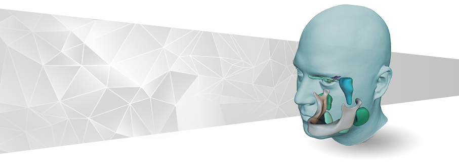
The problem with traditional radiotherapy planning technology is that it is template-based and limited to the use of CT images. Imorphics technology has addressed the challenge and enables automatic contouring for both MRI and Cone Beam CT (CBCT) images.
While CT still offers excellent spatial resolution, high geometric integrity, short exam times, and accurate electron density information to enable dose calculation, the use of MRI in radiotherapy planning is being rapidly adopted due to it’s exquisite soft tissue contrast and visualization. Imorphics’ accurate auto-contouring of MR images without an associated CT image, increases the utility of MR-based radiotherapy planning enormously.
 In addition, image-guided radiotherapy (IGRT) increasingly uses CBCT to ensure that organs at risk are protected during multiple radiotherapy sessions. CBCT offers rapid anatomical visualization with a low radiation dose but the acquired images are low contrast and reconstruction artefacts makes the usual atlas-based approach to auto-contouring fail. Imorphics statistical modelling technology produces excellent auto-contouring results, even when working with missing or spurious data by providing an accurate estimate of the probable anatomy in the image.
Find out more about the Imorphics advantage in radiotherapy planning. Get in touch to talk about how we can help or read about our award-winning MR image segmentation at the PROMISE12 Prostate Grand Challenge and CT image segmentation at the 2015 Head & Neck Grand Challenge.
.
